Treatment of cerebral arteriovenous malformation hemangioma (Previous)
Cerebral Arteriovenous Malformation (AVM) is a congenital cerebrovascular lesion that affects a wide range of age groups, from children as young as 4 years old to elderly people in their 80s. If a congenital AVM ruptures, it can lead to a severe hemorrhagic stroke, which has a great impact on the patient’s neurological function and physical and mental disabilities, and also puts a heavy physical, mental and financial burden on the family.
Causes of cerebral arteriovenous malformation hemangioma
Cerebral arteriovenous malformation hemangioma is a congenital disease with no known cause and no genetic predisposition. The problem of AVMs develops in the mother’s brain during fetal development and is a malformation of the blood vessels somewhere in the brain. The malformed tissue lacks the normal microfilamentous vascular system that allows the arterial and venous blood vessels in the brain to connect directly to each other.
Normal blood supply to the human brain comes from the large blood vessels of the heart and neck, and then through the four aortic vessels of the brain, through the branching blood vessels to the tiny microfilamentary vascular system. Oxygen and nutrients in the blood will permeate through the walls of the microfilamentary blood vessels to nourish the brain cells, and then the blood will be drained through the venous system and return to the veins in the neck, and then flow back to the heart to complete the cycle.
In addition to providing oxygen and nutrients to the brain cells, another function of the microfilamentous blood vessels is to cushion the pressure of the blood flowing into the arteries. However, the center of congenital arteriovenous malformation angiomas lacks the normal microfilamentous vascular system, leaving the veins without a cushion to withstand the immense pressure of arterial blood, causing the veins to become distended and their walls to become fragile, making them susceptible to rupture and hemorrhage, which can then lead to hemorrhagic strokes.
Gender, age distribution and symptoms of patients with cerebral arteriovenous malformation hemangiomas:
Asymptomatic: The majority of patients are asymptomatic from birth until the time the malformation bursts.
Most patients are asymptomatic from birth until their malignant hemangioma bursts.-
Symptoms of unstable blood supply to the brain.
Malformed blood vessels can reduce the proportion of oxygen to brain cells in their vicinity, leading to neurological symptoms such as occasional slurred speech, limb paralysis, unstable vision, and even epileptic seizures. -
Hemorrhagic Stroke
Congenital cerebral arteriovenous malformation hemangiomas (CAVMs) are more common in females and occur in a wide range of ages, from children as young as 4 years old to those in their 80s. Although the risk of cerebral artery malformation hemangioma is less than 1% per year, it is one of the leading causes of hemorrhagic stroke in children, young adults, pregnant women, and healthy individuals, mostly in their late teens to early 40s. People with congenital cerebral arteriovenous malformation (CAVM) hemangiomas usually grow up without any symptoms until the hemangioma bursts, when they experience severe headache, dizziness, nausea, vomiting, neurological disorders, confusion, seizures, and even coma.

Diagnosis of cerebral arteriovenous malformation hemangioma
Since patients usually do not have any symptoms from childhood until the hemangioma bursts, it is advisable for anyone to have a detailed physical examination of the brain and its vascular structure even if they do not have any symptoms, so as to diagnose congenital or acquired cerebrovascular pathology in advance, so as to plan for a response to the hidden time bomb in the brain before it bursts and causes a serious hemorrhagic stroke. It is only by doing so that we can plan our response and choose treatment options before the hidden time bomb explodes in the brain and causes a severe hemorrhagic stroke. With preventive treatment, many tragedies and regrets can be greatly minimized.
The following options are available for a detailed structural examination.
Magnetic Resonance Imaging (MRI) and Magnetic Resonance Angiography (MRA)
Magnetic Res onance Imaging (MRI) is a “non-radiation, non-invasive, and painless” scanning technique, which is very different from X-ray or Computed Tomography (CT scan) scan ning, which carries radiation. The advanced MRI machine not only provides 3D 3D cerebral angiography without the need for contrast injection, but also can accurately and clearly visualize the location of AVMs and their relationship to normal brain tissue. In addition, MRI can scan different parts of the body, providing a thorough screening of the whole body for people with no symptoms, and detecting whether there is a potential risk of stroke or cancer in the body structure.

Computed Tomography Angiography (CTA)
Computed tomography angiography (CTA), which involves radiation and intravenous injection of contrast material, is not the imaging test of choice for general health screening, but it can provide additional information that neurovascular surgeons can use when choosing medical options.
Dynamic and 3D 3D Digital Subtraction Angiography (DSA)
Although minimally invasive and involving radiation and contrast injection, DSA is the gold standard final test required by neurovascular surgeons before selecting a treatment plan. The dynamic flow and velocity of blood from arteries, to microfilaments, to veins, allows doctors to have a complete picture of the blood vessels in the brain. In addition, DSA can help doctors determine whether a malformed vessel is at risk of bursting within a short period of time, so that they can determine the optimal medical treatment plan and timeframe for the patient.
Common Clinical Treatment Options
Current treatment options include the following:
Conservative Observations
If the patient is completely asymptomatic, or if he or she is symptomatic but is elderly, in severely poor health, or if the doctor believes that any treatment would be more risky than no treatment at all in the particular case, conservative treatment is usually observed.
Minimally Invasive Microscopic Excision (MICRO)
The surgery is performed under 3D computerized navigation, and the neurovascular surgeon will also use motor neurocortical reflexes and continuous brain function monitoring system if necessary. Under the microscope, in addition to removing the arteriovenous malformation and removing the hematoma, every effort is made to protect the patient’s neurological function in the brain from damage. The success rate of the surgery depends on the size, location and complexity of the AVM, as well as the experience of the surgeon in charge.
Minimally Invasive Endovascular Embolization (MIE)
Depending on the clinical situation, catheter occlusion surgical therapy can be performed independently, prior to microscopic surgical resection or prior to radiation therapy to reduce the size and extent of the AVM. Neurovascular surgeons use tiny catheters from the arteries in the groin of the patient’s thighs to travel through the large arteries of the patient’s body, navigating the catheters to the vessels of the patient’s brain under X-ray guidance, and then injecting tiny titanium wires, plastic beads, or special glue into the AVMs to embolize the arteries that feed the AVMs and the centers of the AVMs. The main risk of this procedure is accidental injury to both normal and malformed blood vessels during the procedure, causing them to block or rupture and bleed, resulting in an ischemic or hemorrhagic stroke. Another risk of the procedure is the inadvertent premature blockage of the vein responsible for draining blood from the malformed vessel, causing the blood to be drained from the malformed vessel and the pressure in the blood to build up so rapidly that the aneurysm bursts.
Radiotherapy (Radiosurgery)
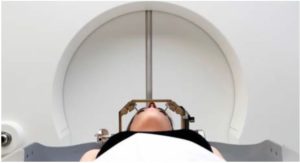
Radiation therapy does not require anesthesia. The treatment involves the delivery of radiation from multiple angles, and the high-energy radiation will be focused on the malformed blood vessels, causing the abnormal malformed blood vessels to slowly and progressively occlude over a period of two to three years, making it impossible for blood to flow through the malformed blood vessels back to the normal blood vessels. With the disappearance of the malformation, the patient’s risk of hemorrhagic stroke is greatly reduced. Radiation therapy is indicated when the size of the malformed hemangioma is less than three centimeters, when the hemangioma is too deep in the brain, when the hemangioma is too close to vital nerve functions, or when the vascular surgeon has assessed that there is a high risk of vascular occlusion or microscopic resection.
The risk of radiation therapy is that although there is no need for anesthesia or surgery, radiation therapy involves radiation, and the radiation must pass through normal brain tissue to reach the malignant hemangioma, which will inevitably cause short-term or long-term effects on the nearby brain cells and vascular tissues. In addition, if the location of the radiation treatment is not precise enough, it may cause premature occlusion of the venous portion of the malformed blood vessel. At this time, blood will only flow into the malformed blood vessel through the arterial blood vessel, but will not be able to flow out through the occluded venous blood vessel, which will increase the blood pressure in the malformed blood vessel and burst, leading to a hemorrhagic stroke.

The system options for radiation therapy are X-ray Knife (X-Knife), Gamma Knife (Gamma Knife), and Digital Guided Knife (Cyberknife).

Congenital cerebral arteriovenous malformation hemangiomas are more common in females and cover a wide range of ages, from children as young as 4 years old to the elderly in their 80s.


